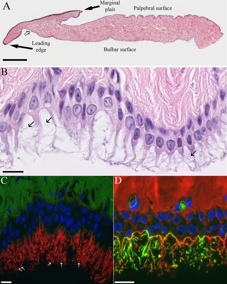
Fig 6. Light microscopic views of the third eyelid.
(A) Panoramic composite view of H&E-stained paraffin section of third eyelid made from 14 images. The point where the smooth stratified epithelium on the bulbar surface first begins to show cytoplasmic extensions is marked by the open arrow. Bar = 500 μm. (B) Bulbar surface of the third eyelid illustrating cytoplasmic extensions with lateral cytofilia (arrows) coming off of them. Bar = 10 μm. (C) The apical membrane near the base of the cytoplasmic extensions is only labeled by MAL-I (green) while the membrane of the extensions and cytofilia are intensely labeled by ECL (red) Arrows point to circular profiles of the bulbous ends of the cytofilia. Bar = 10 μm. (D) Even when a cocktail of WGA, SBA, DBA, PNA and SBA (all red) are used, some cytofilia are more strongly labeled by GSL-I (green). The relative absence of lectin-stained secretory granules in Figs 6C and 6D underscores the negligible contribution of this epithelium to the mucin component of the tear film. Bar = 10 μm.

