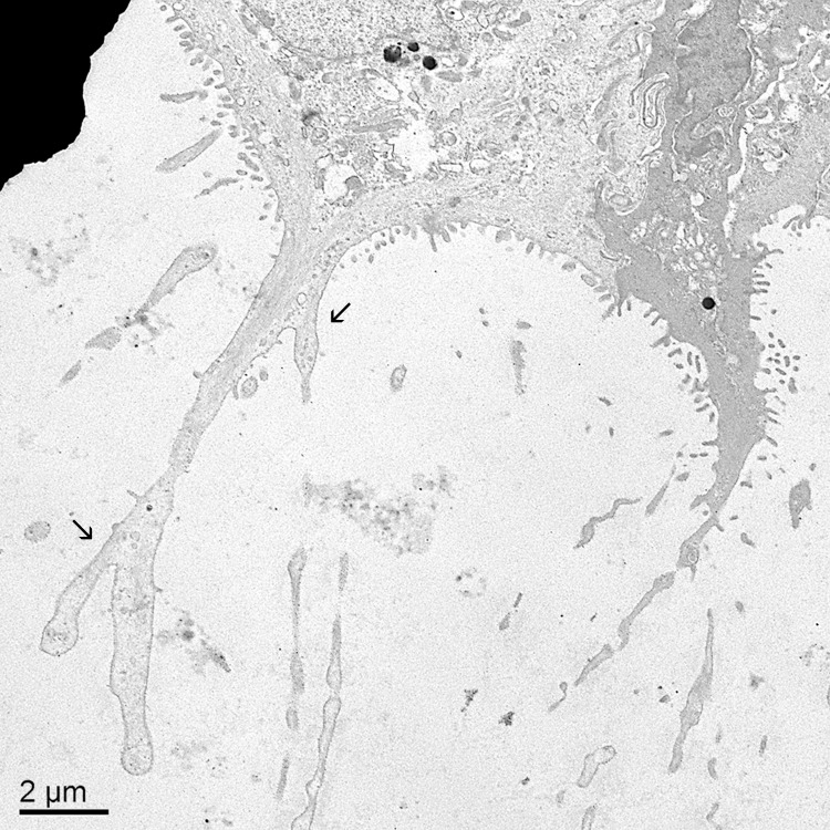
Fig 8. Electron microscopic view of branching, irregular cytoplasmic extensions and cytofilia.
The diameter of the cytoplasmic extensions and cytofilia were often irregular. Cytofilia frequently branched (arrows). Note the relative absence of secretory vesicles in the cytoplasmic extensions. Bar = 2 μm.

