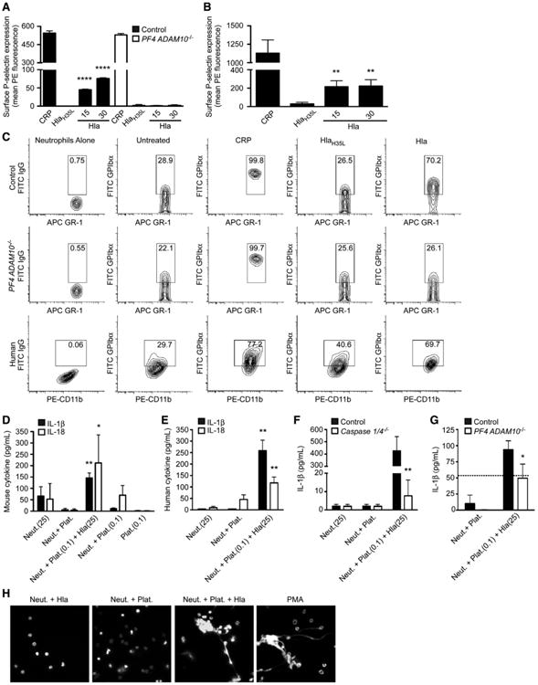
Figure 3. Hla Promotes PNA Formation.

(A and B) P-selectin expression on mouse (A) and human (B) platelets following treatment with collagen-related peptide (CRP), HlaH35L, or Hla; **p ≤ 0.01, ****p ≤ 0.0001.
(C) PNA formation from control and PF4 ADAM10−/− mice (GR1+/GPIbα+) or humans (CD11b+/GPIbα+) either left untreated or treated with CRP, HlaH35L, or Hla.
(D–G) IL-1β (βλαχκ) and IL-18 (white) production following PNA formation in platelet-neutrophil suspensions from wild-type mice (D), human (E), control and Caspase 1/4−/− mice (F), or control and PF4 ADAM10−/− mice (G); (D) and (E) *p ≤ 0.05, **p ≤ 0.01 compared to Neut. or Neut.+Plat. (F) and (G) *p ≤ 0.05, **p ≤ 0.01 compared to control, where dotted line denotes the response of control neutrophils to Hla. Data are represented as mean ± SD. In (D)–(G), (0.1) and (25) denote treatment with toxin concentrations in μg/ml.
(H) NET release examined in mouse platelet-neutrophil suspensions treated as in (D)–(G) or with phorbol myristate acetate (PMA). See also Figure S3.
