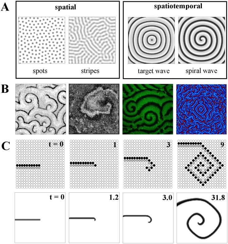
Fig 1. Introduction to pattern types and spiral wave formation.
A: (Left to right) spatial and spatiotemporal pattern examples. Spot and stripe Turing patterns both in coupled Schnakenberg elements; target wave formation from a central pacemaker and established spiral wave, both in coupled FitzHugh-Nagumo oscillators. B: Snapshots of spiral wave patterns from diverse biological systems: (left to right) cAMP signaling in a Dictyostelium discoideum colony, local contraction in neonatal rat cardiac monolayer cultures, MinD protein density in a lipid bilayer and simulated cytokine levels in a two-dimensional grid of cells. See Acknowledgments for image sources. C (upper row): The update rules of the minimal three-state cellular automaton model lead to spiral wave formation, when applied to an open wave front (consisting of a layer of excited cells, depicted in black, and an adjacent layer of refractory cells, depicted in gray). Lower row: a similar numerical experiment for the model from [6,7].

