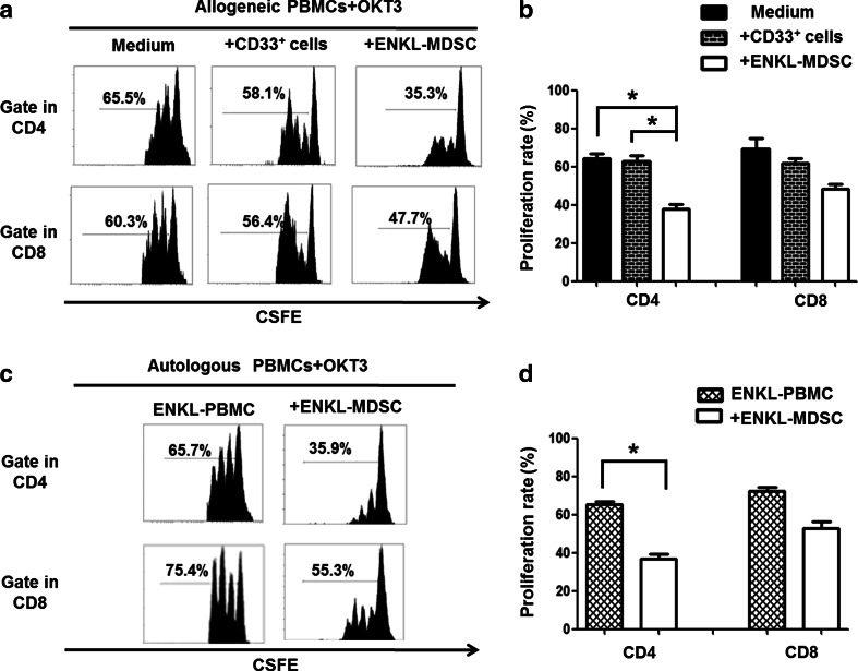
Fig. 3.
ENKL-MDSCs suppress allogeneic and autologous T cell proliferation. T cell proliferation is examined by CSFE labeling in vitro. The CD33+ cells are sorted from the PBMCs from five patients with ENKL, and CD33+ cells from healthy donors are included as a control. The CSFE-labeled PBMCs are co-cultured with the CD33+ cells at a ratio of 2:1 in OKT3-coated 96-well plates. After 3 days, the cells are collected and quantified using flow cytometry. a, c Allogeneic and autologous OKT3-stimulated PBMCs. Representative FACS density plots from one of the five experiments. b, d The graph of the statistical analyses is presented. The error bars represent the SEM. n = 5; *P < 0.05; HD healthy donors

