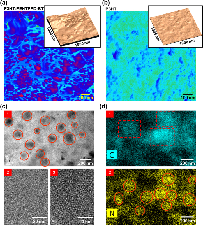
Figure 6. BHJ nanomorphology.
(a,b) Phase-mode AFM images (1 μm × 1 μm) for the P3HT:PEHTPPD-BT layer (a) and the pristine P3HT layer (b), which were coated on the PVP-MMF/ITO-glass substrates (insets: height-mode images). (c) TEM images for the P3HT:PEHTPPD-BT layer (film) (1: low magnification image; 2: bright part image; 3: dark part image): Note that the ‘2’ and ‘3’ images were measured separated by focusing on each part after increasing the magnification of HRTEM system. (d) STEM images for chemical mapping of carbon (1) and nitrogen (2) atoms in the P3HT:PEHTPPD-BT layer (film): Note that the atom distribution is quite broad because of the transmission mode in the STEM measurement which delivers the whole atom information in the thickness direction.

