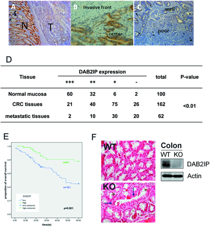
Figure 1. Loss of DAB2IP facilitated the tumor progress in CRC patients.

(A–C) Immunostained comparison of DAB2IP in adjacent normal mucosa and primary site of tumor (A); center and invasive front of tumor (B); well and poor differentiation (C) (magnification: 100x). (D) Expressions of DAB2IP in CRC tissues, lymphatic metastatic tissues and adjacent normal mucosa. (E) Kaplan-Meier overall survival analysis on colorectal patients classified by total DAB2IP expression in primary site; n = 61 for DAB2IP high group, n = 101 for DAB2IP low group. The ‘censored’ means the researchers does not get the precise survival data because of loss follow-up, accidental death and other reasons. (F) Focal aberrant dilated crypt (arrows) in the colonic mucosa from a KO subject (H&E, magnification: 200x).
