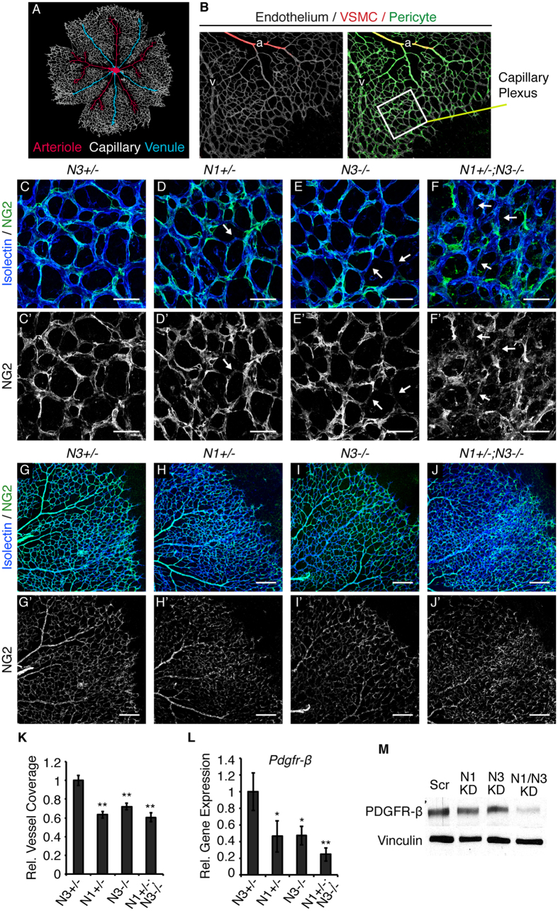
Figure 2. Notch deficiency impairs pericyte coverage.
(A) Schematic of the vascular plexus of a P5 mouse retina with red arterioles, blue venules, and capillaries shown in grey. (B) P5 retina whole-mount stained for isolectin B4 (grey) to identify the endothelium. α-SMA (red) is restricted to VSMCs lining arterioles. NG2 (green) identifies pericytes on capillaries and venules and α-SMA-positive VSMCs. Area enclosed by white box represents an example of a region used for assessment of the capillary plexus. (C–F) P5 retinas whole-mount immunostained for isolectin B4 (blue) to mark endothelial cells and NG2 (green) to mark pericytes. (C’–F’) Corresponding NG2 staining in grey. White arrows mark endothelium devoid of pericyte coverage. (G–J) Example images used for quantification of pericyte content of P5 retinal vasculature stained for CD31 (blue) to mark endothelium and NG2 (green) to mark pericytes. (G’–J’) Corresponding NG2 staining in grey. (K) Quantification of vascular pericyte coverage by normalizing NG2 staining to CD31 in P5 retinas, relative to control. (L) Pdgfr-β transcript levels in P5 retinas measured by quantitative real time PCR, normalized to gapdh and relative to control. (M) Western blot for PDGFR-β and vinculin (loading control) on HBVP protein lysates. n≥3; *p < 0.01, **p < 0.0001. Data are mean±s.e.m. Scale bars: (C–F’) 25 μm, (G–J’) 200 μm.

