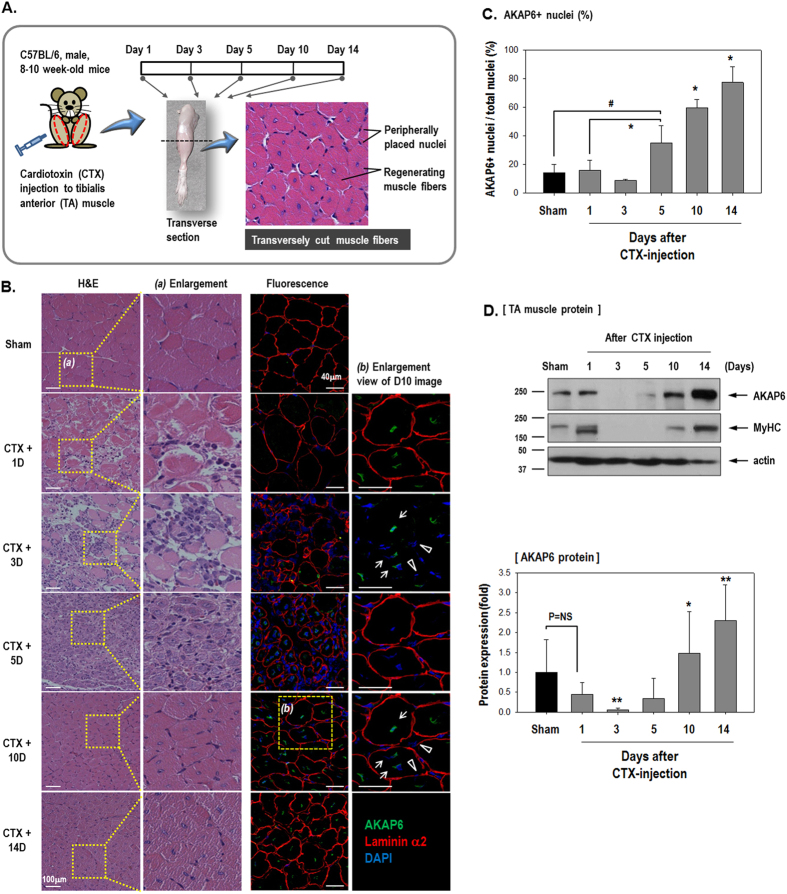
Figure 3. AKAP6 expression during the muscle repair period after injury.
(A) Timeline for the in vivo experiment. (B) Hematoxylin and eosin (H&E) or immunofluorescence staining of transverse muscle sections after cardiotoxin (CTX) injection to the tibialis anterior (TA) muscle on the indicated days. Sham sections were from PBS-injected muscles. H&E staining (Magnification: ×200). Immunofluorescence staining shows that AKAP6 (Green) is predominantly observed in a centrally located nuclear envelope (arrows). Nuclei in a peripheral position in mature myofibers did not express AKAP6 (arrowheads). Laminin α2 for muscle fiber (Red), DAPI for nuclei (Blue; magnification: ×400). Immunofluorescence staining was performed in four mice using four different sections per mouse. (C) The graph shows the percentage of AKAP6-expressing nuclei divided by the number of nuclei (n = 5 each). #p < 0.05 versus Sham; *p < 0.05 versus Day1 CTX (one-way ANOVA). (D) At the indicated time points, the TA muscles were frozen directly in liquid nitrogen, lysed in RIPA buffer, and analyzed with western blotting (Top). Quantification graph of the AKAP6 western blot shows fold changes from values in sham mice (Bottom, n = 4). *p < 0.05 and **p < 0.01 versus Day1 CTX (one-way ANOVA).

