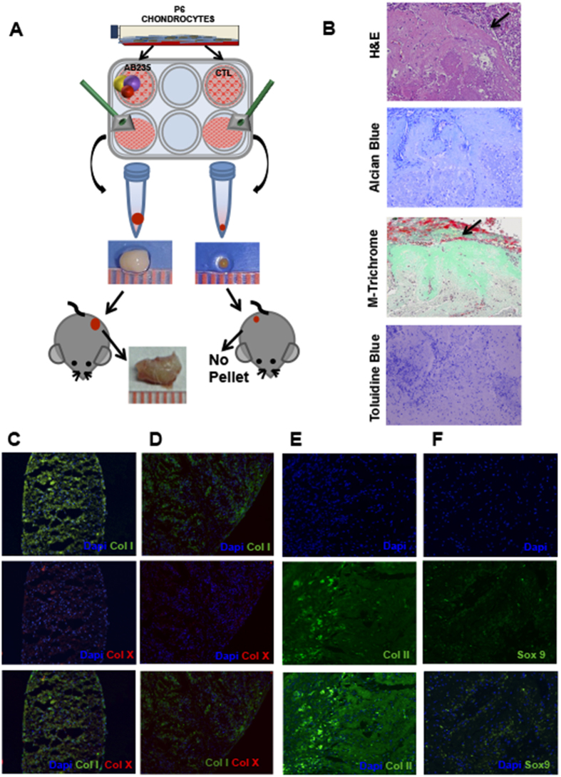
Figure 4. AB235 induces chondrocyte redifferentiation in vivo.
(A) Schematic representation of the experimental design showing images of representative pellets before and after implantation into mice and integration of AB235-treated pellets with the surrounding tissue. (B) Sections of AB235-treated pellets harvested from mice and stained for H&E, Masson’s Trichrome, Alcian Blue and Toluidine Blue show a robust staining for mature, cartilage-like ECM. Black arrows indicate the edge of the pellet in H&E and Masson’s Trichrome stained sections while the edge of the pellet is not visible in Alcian Blue and Toluidine Blue stained sections. (C–F) Representative images of immunofluorescence analysis of cartilage markers. Stained sections of fibrotic marker type I collagen (Col I) and hypertrophic marker type X collagen (Col X) in both control pellet grown in vitro (C) and AB235-induced pellet harvested from mice (D). Expression of the chondrogenic markers Col II and Sox 9 in AB235 induced pellet sections after the in vivo assay (E,F). Original magnification 10× for (C,D); 20× for (E,F).

