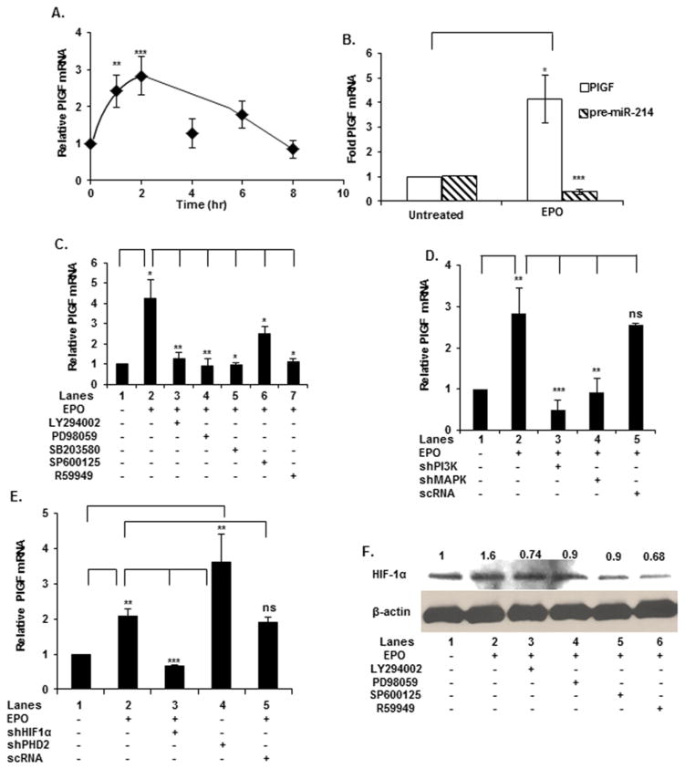
Figure 1. EPO-induced PlGF mRNA expression involved signalling via the PI3 kinase, MAP kinase, JNK and HIF-1α pathways.
(A) K562 cells were treated with EPO for indicated time periods followed by isolation of total RNA for qRT-PCR analysis of PlGF mRNA. (B) CFU-E cells were treated with EPO for 2 h followed by isolation of RNA and qRT-PCR analysis. (C) K562 cells were pre-treated for 30 min with the indicated inhibitors followed by EPO-treatment for 2 h. (D) K562 cells were transfected, as indicated, with 1 μg of shRNAs for PI-3 kinase, MAP kinase and scRNA followed by EPO treatment for 2 h. (E) Cells were transfected, as indicated, with 1 μg of shRNAs for the transcription factor HIF-1α, scRNA and PHD2 followed by treatment with EPO for 2 h. In panels (A–D) total RNA was subjected to quantitative qRT-PCR for PlGF mRNA. PlGF mRNA expression was normalized to GAPDH mRNA levels. Data are expressed as means ± S.D. of three independent experiments. (F) Western Blot analysis of HIF-1α protein in K562 cells treated with the indicated pharmacological inhibitors followed by EPO treatment for 2 h. All Western blots were stripped and reprobed with β-actin to normalize protein loading. Data are representative of three independent experiments. ***P < 0.001, **P < 0.005, *P < 0.01, ns, P > 0.05.

