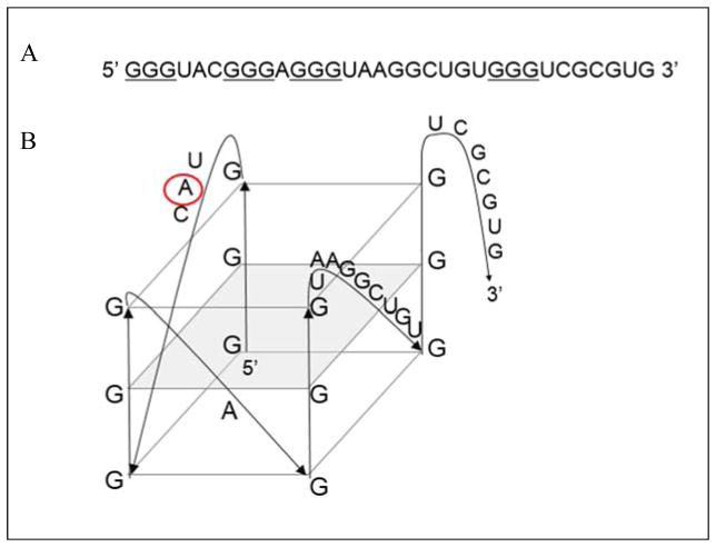
Figure 1.
(A) 32 nt guanine rich sequence located in the 3′-UTR of NR2B mRNA (NM_000834.3, position 4659) predicted to form G quadruplex (guanine triplets proposed to be involved in the structure are underlined). (B) Arrangement of the predicted G quadruplex structure in the NR2B mRNA. QGRS Mapper software was used for the prediction (http://bioinformatics.ramapo.edu/QGRS/analyze.php)42. Adenine replaced by the 2-amino purine fluorescent analog is circled in red.

