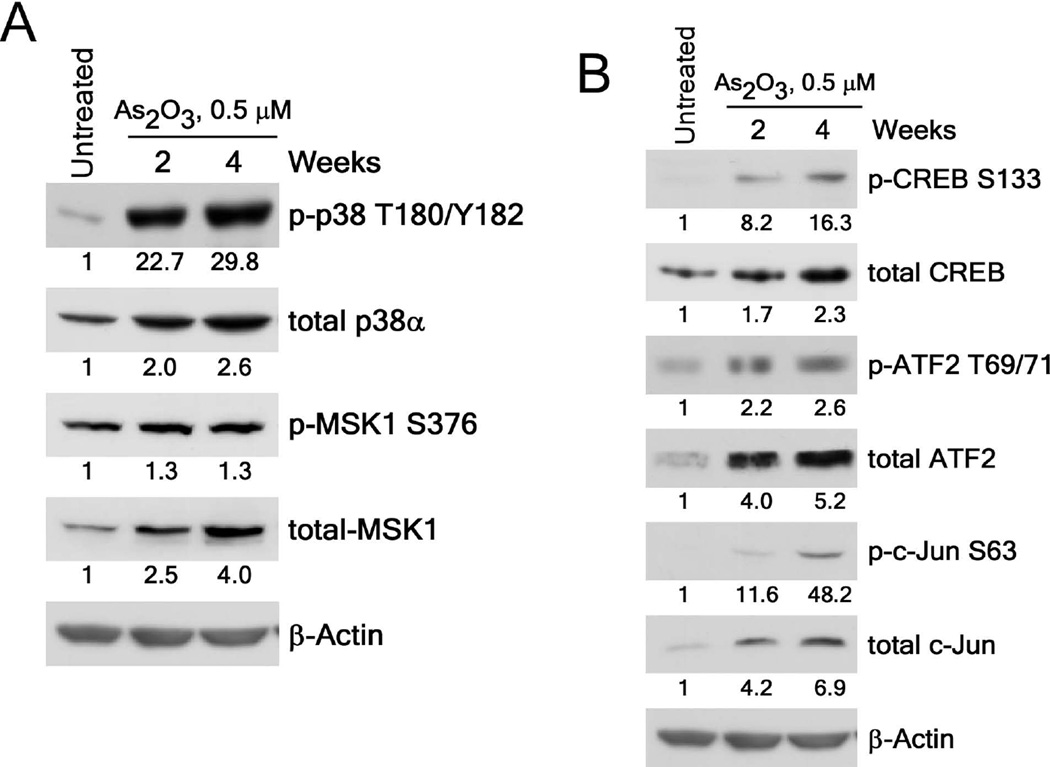
Figure 2.
Arsenic exposure activates the p38α-signaling cascade in BALB/c 3T3 cells. (A) Phosphorylation and expression of p38α are increased in arsenic-treated BALB/c 3T3 cells. (B) Arsenic increases the phosphorylation and expression levels of the CREB, ATF2 and c-Jun transcription factors in BALB/c 3T3 cell lysates. Numbering indicates the fold increase compared to untreated control as 1. Cellular proteins were resolved by 10% SDS-PAGE and the protein levels were visualized by Western blotting with specific primary antibodies and a horseradish peroxidase (HRP)-conjugated secondary antibody. Detection of total β-actin was used to verify equal protein loading.

