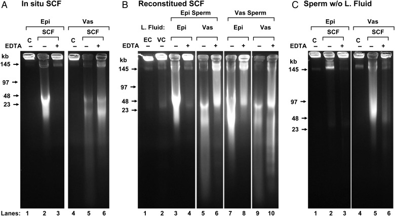
Figure 1.
FIGE analysis of SCF. (A) In situ sperm chromatin fragmentation (SCF): epididymal (lanes 1–3) and vas deferens (lanes 4–6 sperm were induced to undergo SCF by incubation with Mn2+ and Ca2+ in the presence of their luminal fluid, without (lanes 2 and 5) or with subsequent EDTA treatment to reverse the double-stranded DNA breaks (dsDSBs) (lanes 3 and 6). Lanes 1–6 are reproduced from Yamauchi et al. (2007a). Control, untreated samples are shown in lanes 1 and 4 (B) reconstituted SCF: epididymal sperm were washed and resuspended in luminal fluid from the epididymis (lanes 3 and 4) or vas deferens (lanes 5 and 6) then induced to undergo SCF, without (lanes 3 and 5) or with subsequent EDTA (lanes 4 and 6). Vas deferens sperm were resuspended in luminal fluid from the epididymis (lanes 7 and 8) or vas deferens (lanes 9 and 10) then induced to undergo SCF, without (lanes 7 and 9) or with subsequent EDTA (lanes 8 and 10). Control, washed sperm from the epididymis (lane 1) and vas deferens (lane 2). (C) Sperm-induced to undergo SCF without luminal fluid: epididymal (lanes 1–3) and vas deferens (lanes 4–6) sperm were induced to undergo SCF without (lanes 2 and 5) or with (lanes 3 and 6) subsequent EDTA treatment. For each lane in the gel, two experiments were performed using one mouse each. Only one experiment is shown.

