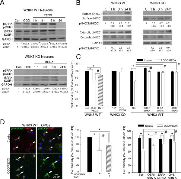
Figure 5. Knockout of WNK3 or SPAK/OSR1 protects neurons and OPCs against ischemic damage.
A. Representative immunoblot of pSPAK and pOSR1 expression in primary cortical neurons of WT or WNK3 KO mice after 2 h oxygen-and-glucose deprivation (OGD) or after subsequent reoxygenation (REOX) for durations of 1, 3, 6 or 24 h. B. Neuronal expression of pNKCC1 in cytosolic fraction or at the cell surface membrane as detected by surface biotinylation (see Supplemental Methods). Values represent Mean ± SEM (n = 3-5). C. Summary of neuronal viability data under control or Scrambled (Scr) control conditions, OGD/REOX plus bumetanide (BMT; 10 μM for 24 h), or treated with SPAK and OSR1 siRNAs. Data are normalized to the scrambled group. Values represent Mean ± S.E.M. (n = 3). *P <0.05. # P < 0.05. D. Elevated expression of pSPAK/OSR1 and pNKCC1 in MBP-positive OPCs after OGD/REOX. Right panels, OPC survival after OGD/REOX with NKCC1 inhibition by BMT. Knockdown of SPAK/OSR1 by siRNA increases viability of OPC. Values represent Mean ± S.E.M. (n = 3-5). *P < 0.05. # P < 0.05.

