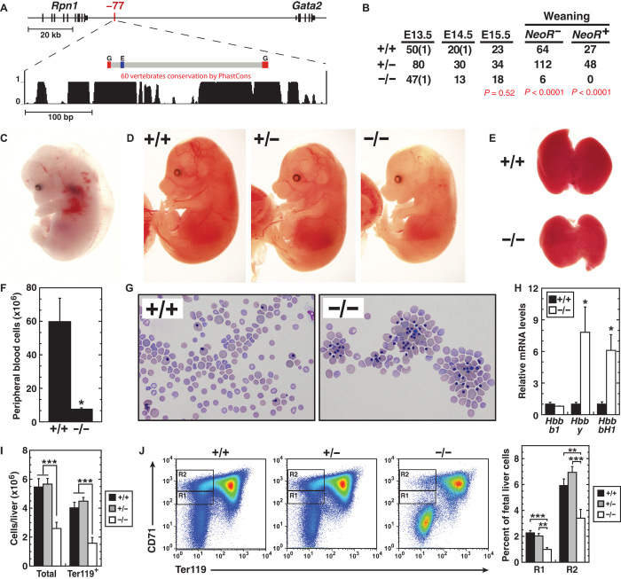
Fig. 1. −77 leukemogenic, long-range enhancer is essential for embryogenesis and hematopoiesis.
(A) Vertebrate conservation plot of −77 showing positions of GATA motifs (G; WGATAR) and E-box (E; CANNTG) within the deleted region. (B) Genotypes of −77+/+, −77+/−, and −77−/− NeoR- embryos at timed developmental stages and genotypes of NeoR− and NeoR+ pups at time of weaning. (C) Representative E13.5 +9.5−/− embryo exhibiting anemia, hemorrhage, and edema. (D) Representative E15.5 −77+/+, −77+/−, and −77−/− littermates. (E) Representative E15.5 fetal livers from −77+/+ and −77−/− littermates. (F and G) E15.5 peripheral blood quantitation [−77+/+ (n = 3) and −77−/− (n = 3)] and representative Wright-Giemsa staining. (H) Relative expression of fetal and adult β-globin mRNA in peripheral blood at E15.5. (I) Total cells [−77+/+ (n = 10), −77+/− (n = 24), and −77−/− (n = 13)] and Ter119+ cells [−77+/+ (n = 7), −77+/− (n = 17), and −77−/− (n = 5)] in E13.5 fetal livers. (J) Representative flow cytometric analysis and quantitation of E13.5 fetal livers for CD71+Ter119− R1 and R2 erythroid progenitors [−77+/+ (n = 7), −77+/− (n = 17), and −77−/− (n = 5)]. Graphs show means ± SEM; *P < 0.05, **P < 0.01, ***P < 0.001.

