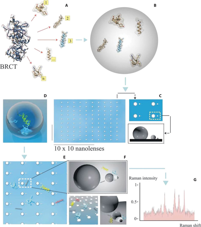
Fig. 1. Scheme of the whole process from peptide extraction to Raman detection.

(A) A mixture of peptides is extracted from the BRCT domain derived from the BRCA1 protein. (B) The mixture is collected in aqueous solution at a concentration of 100 pM. (C to E) A drop is deposited and dried on a matrix array of SSCs (each chain is a pixel). (F) The whole content of peptides is spread over the matrix, and some (three different types per pixel, on average) are deposited in the smallest gap. These are the only ensembles per pixel that contribute to the Raman signal. (G) Representative Raman signal from pixel i,j of the matrix array composed of, on average, three different types of peptides.
