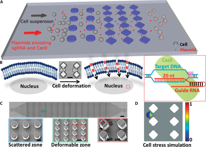
Fig. 1. Delivery mechanism and device design.

(A) Illustration of the delivery process wherein cells pass through the microconstriction and experience deformability. Plasmids encoding sgRNA and Cas9 protein are mixed with the cells to flow through the chip. (B) Illustration of the delivery mechanism whereby transient membrane holes are generated when cells pass through the microconstriction and specific genome editing is conducted after plasmids encoding sgRNA and Cas9 protein are delivered into the cell. Cell deformation was shown by microscopy when cells passed through the microconstriction. Scale bar, 15 μm. nt, nucleotide. (C) Microscopy of the whole device structure. Scale bar, 0.5 mm. Scanning electron microscopy (SEM) of scattered and deformable zones in the device is also shown. Scale bar, 15 μm. One diamonded microconstriction of 15-μm depth and 4-μm width is indicated by the red arrow. The length of the diamond edge is 10 μm. (D) Cell stress simulation was applied on the diamonded microconstriction design with 15-μm depth and 4-μm width when a cell began to penetrate the constriction. A graphical representation of the cell stress gradient that forms across the membrane is shown.
