
Fig. 3. High-frequency LFP amplitude envelopes are correlated between cortex and SC.
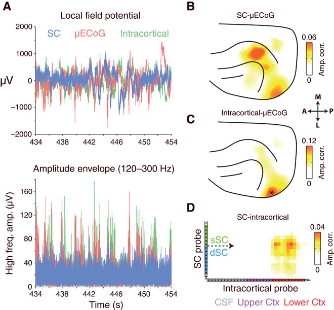
(A) Simultaneously recorded SC, intracortical, and μECoG signals from one example recording session. No relationship between signals is immediately visible upon inspection of LFPs (top). However, high-frequency amplitude envelopes display burst-like fluctuations that appeared to occur synchronously. (B) Cortical topography of high-frequency amplitude envelope correlation for the seed SC electrode displayed in (A). Note the region-specific amplitude correlation for lateral visual and suprasylvian cortical areas. (C) Cortical topography of high-frequency amplitude envelope correlation for the intracortical recording site displayed in (A). Note the presence of correlated high-frequency activity in cortical regions immediately surrounding the position of the intracortical recording site (marked as *). (D) Strength of high-frequency amplitude correlation between all SC and intracortical channel pairs. Note the presence of a cluster of correlated channels in the center of the SC probe and the lower third of the intracortical probe.