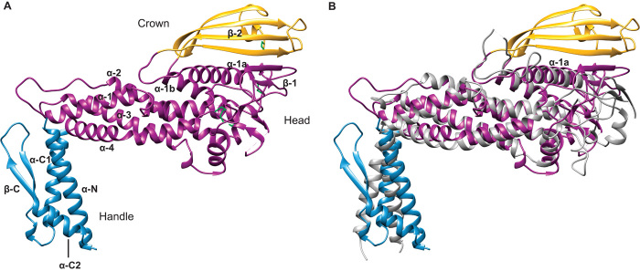
Fig. 1. Comparison of the BabA and SabA extracellular domain crystal structures.
(A) Crystal structure of the BabA extracellular domain. Indicated are the handle (blue) and head regions (dark magenta) and the crown β-strand unit (gold). The four disulfide bridges are represented as green sticks. (B) Superimposition of the extracellular domains of BabA and SabA (gray).

