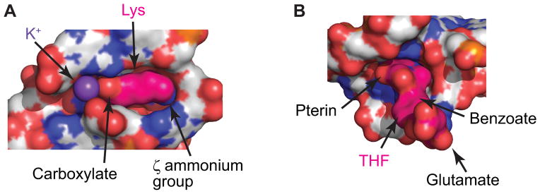
Fig. 4.
Examples of ligand-binding pockets. RNA (grey) and ligands (hot pink) are shown in surface representation with heteroatoms depicted in the following colors: oxygen, red; nitrogen, blue; phosphorus, orange. All views are from the top in respect to the Fig. 3C,D views. (A), Tight lysine-bound pocket of the lysine riboswitch shows good shape complementarity with bound lysine [79]. A potassium cation (violet sphere) mediates interactions between the carboxylate of lysine and RNA. Nucleotides from the front are removed to visualize the pocket. (B) Semi-open pocket of the THF riboswitch bound to THF [90]. The pocket is located in the junctional region.

