
Figure 1.
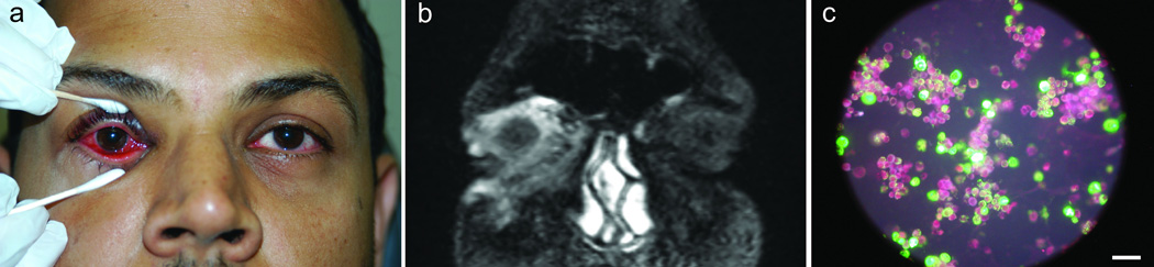
A) Conjunctivitis, right eye. B) Coronal image showing inflammation of the conjunctiva, eyelids, anterior orbit, and pre-malar area. C) Direct fluorescent antibody label (green) against adenovirus antigen, showing punctate cytoplasmic and nuclear staining in A549 cells (red). Scale = 100 microns.