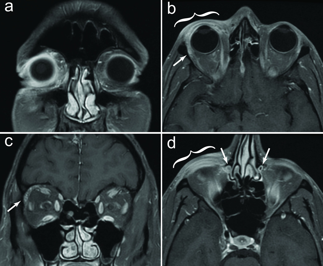
Figure 2.
A) Fat saturation image, demonstrating enhancement of periocular tissues. B) Thickening of right upper eyelid (bracket) and anterior orbital enhancement. Lacrimal gland is enlarged (arrow). C) Coronal image confirming right lacrimal gland enlargement (arrow). D) Inflammation of lower eyelid (bracket), and right nasolacrimal duct swelling with loss of air signal in the lumen (arrows).

