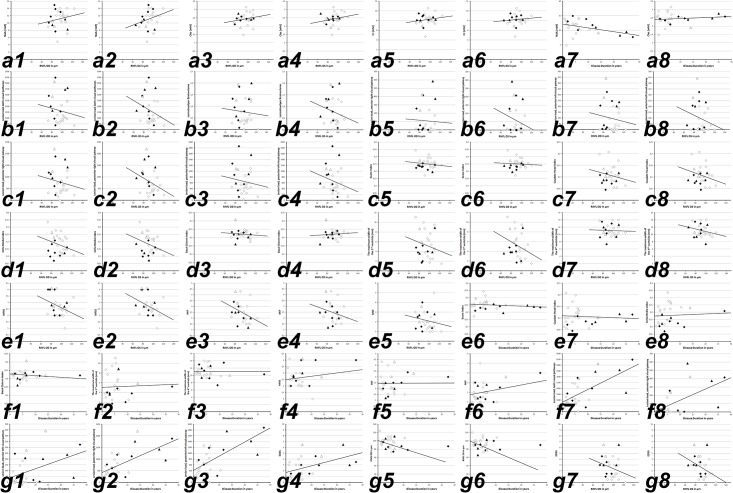
Fig 1.
white squares, RRMS without ON; black squares, RRMS with ON; white triangles, SPMS without ON; black triangles, SPMS with ON; black line, linear regression curve. Abbreviations: OD, oculus dexter (right eye); OS, oculus sinister (left eye); RNFL, retinal nerve fiber layer; NAA, N-acetyl-aspartate; Cho, choline; Cr, creatine; NAWM, normal appearing white matter; MIF, the maximum width of the anterior interhemispheric fissure; MSF, the maximum width of the Sylvian fissure; MFSS, the maximum frontal subarachnoid space; EDSS, expanded disability severity scale. a1-g8, linear regression curves for: a1, RNFL vs. NAA (right eye); a2, RNFL vs. NAA (left eye); a3, RNFL vs. Cho (right eye); a4, RNFL vs. Cho (left eye); a5, RNFL vs. Cr (right eye); a6, RNFL vs. Cr (left eye); a7, disease duration vs. NAA in the NAWM; a8, disease duration vs. Cho; b1, RNFL vs. lesion load (right eye); b2, RNFL vs. lesion load (left eye); b3, RNFL vs. lesion load per brain volume (right eye); b4, RNFL vs. lesion load per brain volume (left eye); b5, RNFL vs. lesion load along anterior right visual pathway (right eye); b6, RNFL vs. lesion load anterior right visual pathway (left eye); b7, RNFL vs. lesion load along anterior left visual pathway (right eye); b8, RNFL vs. lesion load anterior left visual pathway (left eye); c1, RNFL vs. lesion load along posterior right visual (right eye); c2, RNFL vs. lesion load along posterior left visual pathway (left eye); c3, RNFL vs. lesion load along posterior left visual (right eye); c4, RNFL vs. lesion load along posterior left visual pathway (left eye); c5, RNFL vs. Evan’s Index (right eye); c6, RNFL vs. Evan’s Index (left eye); c7, RNFL vs. Caudate Head Index (right eye); c8, RNFL vs. Caudate Head Index Index (left eye); d1, RNFL vs. Cella Media Index (right eye); d2, RNFL vs. Cella Media Index (left eye); d3, RNFL vs. Basal Cistern Index (right eye); d4, RNFL vs. Basal Cistern Index Index (left eye); d5, RNFL vs. the maximum width of the 3rd ventricle (right eye); d6, RNFL vs. the maximum width of the 3rd ventricle (left eye); d7, RNFL vs. the maximum of the 4th width ventricle (right eye); d8, RNFL vs. the maximum of the 4th width ventricle (left eye); e1, RNFL vs. MFSS (right eye); e2, RNFL vs. MFSS (left eye); e3, RNFL vs. MIF (right eye); e4, RNFL vs. MIF (left eye); e5, RNFL vs. MSF (right eye); e6, RNFL vs. MSF (left eye); e7, disease duration vs. Evan’s Index; e8, disease duration vs. Caudate Head Index; f1, disease duration vs. Cella Media Index; f2, disease duration vs. the maximum width of the 3rd ventricle; f3, disease duration vs. the maximum width of the 4th ventricle; f4, disease duration vs. MFSS; f5, disease duration vs. MIF; f6, disease duration vs. MSF; f7, disease duration vs. lesion load along both visual pathways; f8, disease duration vs. lesion load along the anterior right visual pathway; g1, disease duration vs. lesion load along the anterior left visual pathway; g2, disease duration vs. lesion load along the posterior right visual pathway; g3, disease duration vs. lesion load along the posterior left visual pathway; g4, disease duration vs. EDSS; g5, disease duration vs. RNFL (right eye); g6, disease duration vs. RNFL (left eye); g7, RNFL (right eye) vs. EDSS; g8, RNFL (right eye) vs. EDSS. Regression analyses demonstrated only weak correlations between the examined parameters a1-g8 of all 28 MS patients included in this study and associated subgroups (RRMS without ON, RRMS with ON, SPMS without ON, SPMS with ON). Of note, the plotted linear regression curves in a1 –g8 are calculated for the analysis of all included MS patient.

