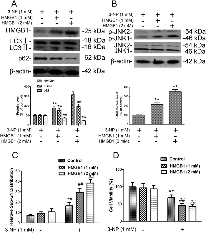
Fig 6. 3-NP–induced expression of HMGB1, p-JNK, LC3, SQSTM1, and apoptosis were increased by exogenous HMGB1 treatment in primary striatal neurons.
(A-B) Cells in culture were exposed to exogenous purified HMGB1 protein (1 or 2 mM) in the culture medium for 24 h followed by 24 h with added 3-NP. Total cellular extracts were subjected to Western blotting for HMGB1, LC3, SQSTM1, and JNK and p-JNK expression analysis. Densities of protein bands were analyzed with an image analyzer (Sigma Scan Pro 5) and normalized to the loading control (β-actin). Bars represent mean ± SE; n = 3 cell samples per group. Groups were compared by ANOVA followed by Dunnet’s post hoc test before data conversion. **p <0.01 vs control (C-D) Treated cells were subjected to cell cycle analysis and cell viability analysis as in Fig 3C and 3D. **p <0.01 vs control. ##p <0.01 vs. control.

