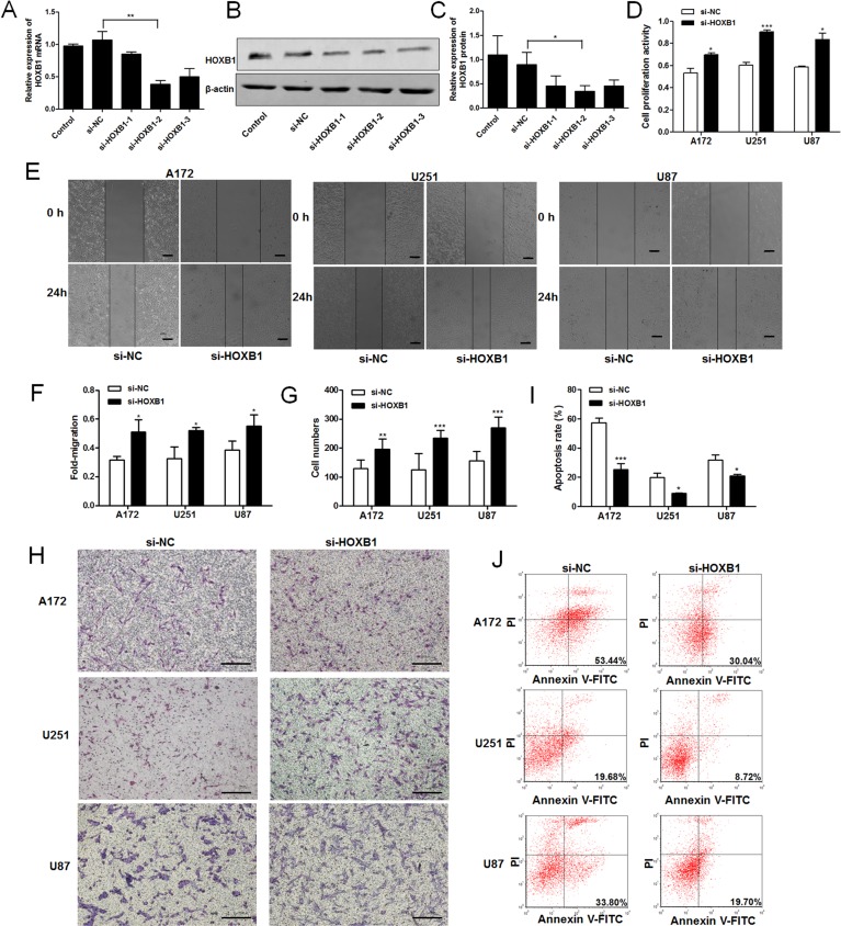
Fig 3. The role of HOXB1 in glioma cell proliferation, invasion, and apoptosis.
(A-C) Three siRNA targeting different sites of HOXB1 and a siRNA negative control (si-NC) were transfected into U87 cells using Lipofectamine 2000. Total mRNA or total protein was isolated at 24 h or 48 h after transfection. HOXB1 mRNA expression was assessed by qRT-PCR. HOXB1 protein expression was determined with western blot analysis. si-HOXB1-2 as the most efficient siRNA was used in further studies. (D) An MTT cell proliferation assay was performed at 48 h after transfection with equal concentrations of si-HOXB1 and si-NC in glioma cell lines. (E-F) Scratch wound assay of glioma cell lines transfected with equal concentrations of si-HOXB1 and si-NC. A linear wound was made with a 200 μl pipette tip and the wound width was recorded at three different reference points after 0 and 24 h (40 ×, Scale bars = 200 μm). (G-H) Transwell assay of glioma cell lines transfected with equal concentrations of si-HOXB1 and si-NC. The migrated cells were fixed with 4% paraformaldehyde, stained with Giemsa and calculated with microscope in three non-overlapping, randomly selected fields (100 ×, Scale bars = 200 μm). (I-J) Flow cytometric analysis of apoptosis in glioma cell lines after transfection with equal concentrations of si-HOXB1 and si-NC. Apoptosis was assessed with Annexin V-FITC/PI. All results are representative of three independent experiments, and a statistical analysis is performed (mean ± SD, *P < 0.05, **P < 0.01, and ***P < 0.001).

