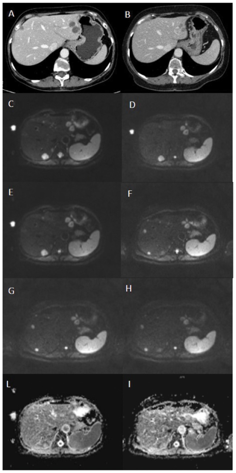
Fig 1. 45-year-old woman with liver metastasis from colorectal cancer.

Portal-phase axial CT scans before (A) and three months after (B) treatment show a decrease in size of two left liver lobe lesions and disappearance of the right liver lobe lesion. Axial DWI images before (C, b value 50, D, b value 400, E, b value 800) and 14 days after treatment (F, b value 50, G, b value 400, H, b value 800) do not demonstrate significant differences in the signal intensity of the two left liver lobe lesions with disappearance of the right liver lobe lesion. Axial tissue diffusion maps before (I) and 14 days after treatment (J) do not show significant differences in the signal intensity of the two left liver lobe lesions with disappearance of the right liver lobe lesion. This demonstrates that the qualitative assessment and the quantitative tissue diffusion-based assessment are not sufficient to evaluate tumor response.
