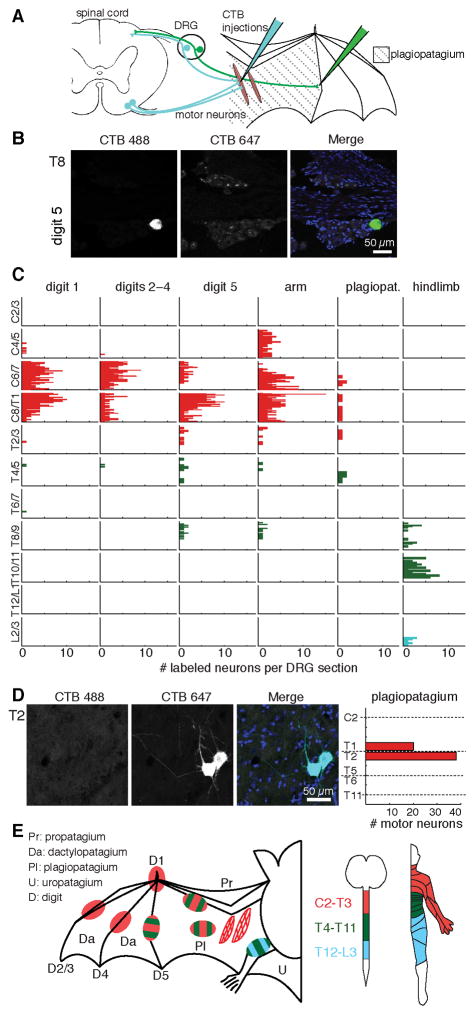
Figure 1. Bat wing neuronal tracing reveals atypical somatosensory-motor innervation.
(A) Schematic of neuronal tracing approach.
(B) T8 DRG section from bat wing injected at digit 5 with CTB Alexa-488 (green). Merged images shows DAPI-stained nuclei (blue).
(C) Histograms show the number of neurons labeled at each spinal level from all injections (≤1.5 μl per injection). Each column shows labeling from a separate wing site (N=2–3 injections per site from 2–3 bats). See also Figure S1. Color key in panel E.
(D) Motor neurons in upper thoracic spinal cord were labeled by injection of CTB Alexa-647 into plagiopatagial muscles. Merged image shows DAPI-stained nuclei (blue). Right, motor neuron quantification (N=6 injections in 2 bats). Dashed lines indicate transection levels of dissected spinal cords (see Supplemental Methods).
(E) Dermatome and myotome maps. Left, injection sites colored according to spinal level of innervation. Motor pools are represented by hatched areas. Middle: spinal level color key. Right, map of corresponding human dermatomes.

