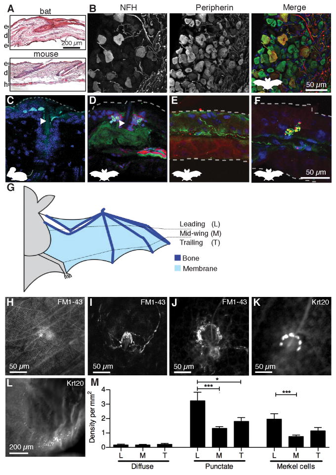
Figure 2. An unusual repertoire of touch receptors innervates bat wings.
(A) Skin histology of bat wing and mouse limb [epidermis (e), dermis (d), hypodermis (h)].
(B) Bat DRG labeled with antibodies against neurofilament H (NFH; red) and peripherin (green). DAPI (blue) labeled nuclei. Labeling and colors apply to B–F. See also Figure S2A.
(C–F) Immunohistochemistry of mouse limb (C) and bat wing skin (D–F). Dashed lines denote skin surfaces. (C) Keratin 8 (Krt8) antibodies (cyan) labeled mouse Merkel cells adjacent to a guard hair (arrowhead). (D) Krt20 antibodies (cyan) labeled bat Merkel cells around a wing hair (arrowhead). (E) Free nerve ending. (F) Knob-like ending. Scale applies to C–F.
(G) Schematic of wing areas.
(H–J) In vivo FM1-43 injections labeled (H) diffuse endings (asterisk) (I) lanceolate endings and (j) sensory neurons similar to mouse Merkel-cell afferents (see also Figure S2B–D).
(K–L) Merkel cells were surveyed using whole-mount Krt20 immunostaining of 12 wing areas (see Figure S2E). Merkel cells were found near hairs (K) and along fingertips (L).
(M) Sensory ending density at wing areas defined in (G). [N=4 wings from four bats (diffuse and punctate), N=4 wings from three bats (Merkel cells)]. Punctate endings and Merkel cells were unevenly distributed across wing areas (One-way ANOVA; P=0.0004 and P=0.002, respectively). Asterisks denote significance between groups by Bonferroni’s multiple comparison test. ***P≤0.001, **P≤0.01, *P≤0.05. Bars: mean ± SEM. See also Figure S2E–H.

