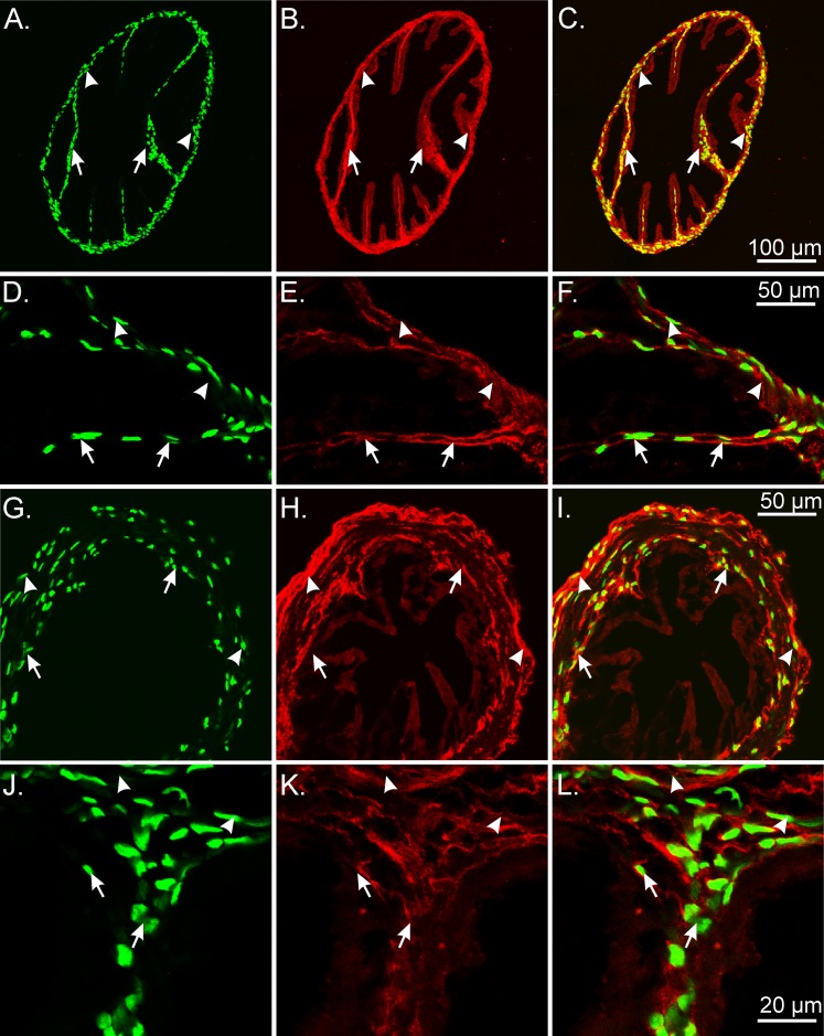
FIG. 3.
The eGFP nuclei within PDGFRα+cells in the mouse oviduct. PDGFRα+ cells were densely distributed within the myosalpinx and endosalpinx. The eGFP nuclei (green) were located within the cytoplasm of PDGFRα+ cells (red) throughout the oviduct. A–F) In the ampulla region, PDGFRα+ cells were detected deep within the endosalpinx folds (arrows), and within the thin-walled myosalpinx (arrowheads). In the isthmus region of the duct (G–I), PDGFRα+ cells were distributed within the epithelial stroma along the inner aspect of the myosalpinx (arrows; red). The cytoplasm of PDGFRα+ cells was also densely distributed on the outer periphery and more loosely distributed within the myosalpinx (arrows). J–L) Higher-power images showing the eGFP nuclei within PDGFR+ cells in the myosalpinx (arrowheads) and endosalpinx folds (arrows). Bars in C, F, I, and L represent their respective series of panels.

