FIG. 6.
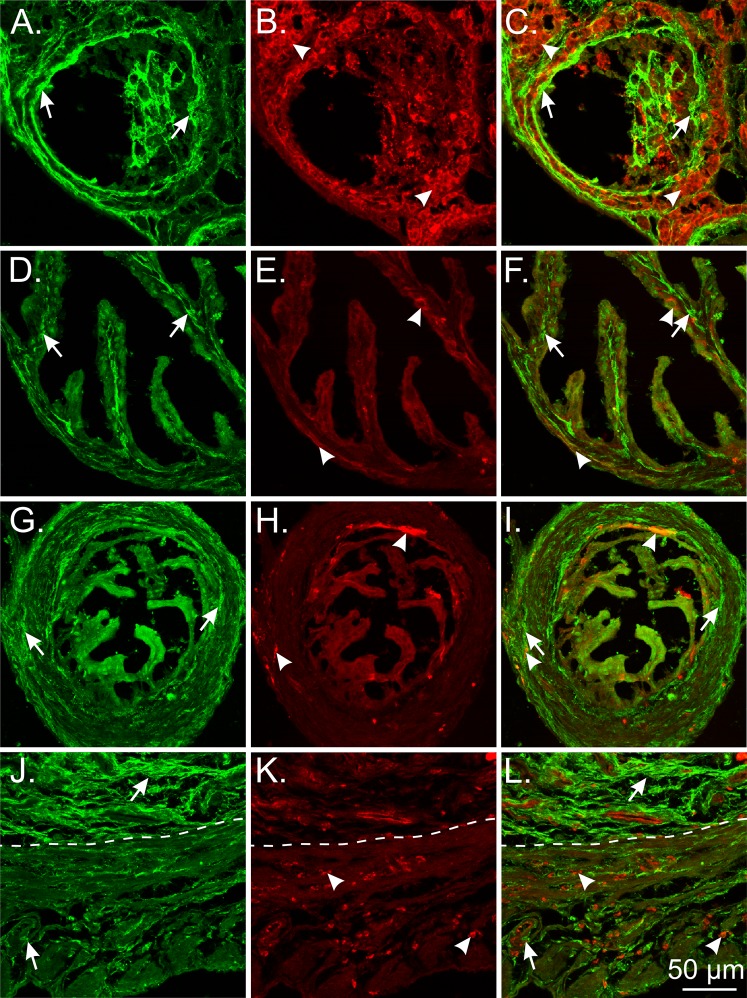
Limited cellular colocalization of PDGFRα and vimentin in the mouse female reproductive tract. A–C) PDGFRα+ cells (green; arrows) and vimentin+ cells (red; arrowheads) in the ovary. Vimentin+ cells surrounded follicles but were distinct from PDGFRα+ cells. D–F) PDGFRα+ cells (green; arrows) and vimentin+ cells (red; arrowheads) in the ampulla region of the oviduct. Few vimentin+ cells were located in the myosalpinx and endosalpinx. G–I) PDGFRα+ and vimentin+ cells in the isthmus region of the oviduct. There was a sparse distribution of vimentin+ cells in the myosalpinx and endosalpinx. J–L) PDGFRα+ cells and vimentin+ cells in the uterus (myometrium and endometrium). Vimentin+ cells were localized around smooth muscle bundles in the myometrium and interspersed between PDGFRα+ cells in the endometrium and within the wall of blood vessels. The dotted line separates the endometrium (upper) from the myometrium (lower). Bar in L applies to all panels.

