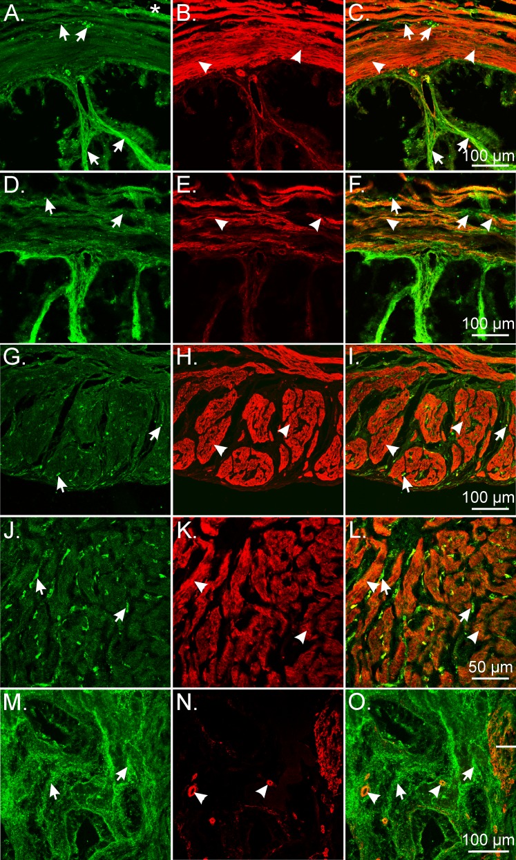
FIG. 8.
Double labeling of PDGFRα and Smmhc reveal distinct populations of cells in the NHP oviduct and uterus. A–F) In the monkey oviduct, PDGFRα+ cells (green; arrows) were located along the serosa (*), within the myosalpinx (arrows), and deeper in the endosalpinx and mucosal folds (arrows) compared with Smmhc+ SMCs (red; arrowheads), which were primarily localized in the myosalpinx and blood vessels. G–L) In the monkey myometrium, PDGFRα+ cells (arrows) were located around muscle bundles (arrowheads) and within septa that separated these bundles. J–L) At higher power, distinct populations of PDGFRα+ and Smmhc+ SMCs are readily distinguished. PDGFRα+ cells within the myometrium (green; arrows) are interspersed between and surrounding smooth muscle bundles (red; arrowheads). M–O) In the uterine endometrium, PDGFRα+ cells formed a meshlike network of cells (arrows) that were distinct from the sparse Smmhc+ SMCs (arrowheads) that were localized to blood vessels. Bars in C, F, I, L, and O represent their respective series of panels.

