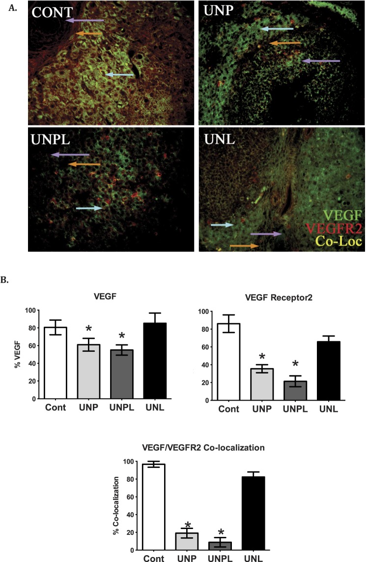
FIG. 3.
Early life undernutrition reduces VEGF, VEGFR2, and their colocalization in offspring ovaries. A) Photographs represent immunopositive staining of VEGF (green), VEGFR2 (red), and the colocalization of VEGF/VEGFR2 (yellow). Negative controls show no positive staining (data not shown). Original magnification ×200. B) Early life undernutrition resulted in a significant decrease in the proportion of VEGF, VEGFR2, and VEGF/VEGFR2 immunostaining (expressed as percent of positive stained area of total ovarian area analyzed) in offspring ovaries. One-way ANOVA main effects: maternal diet P < 0.05. Post hoc analyses (Bonferroni): *P < 0.05 for undernourished compared to control offspring (n = 5 per group). Cont, offspring of mothers fed a control diet; UNP, offspring of mothers undernourished during pregnancy alone; UNPL, offspring of mothers undernourished during pregnancy and lactation; UNL, offspring of mothers undernourished during lactation alone. Cell types are indicated by colored arrows: granulosa cells (purple arrow), theca cells (orange arrow), and ovarian stroma (light blue arrow).

