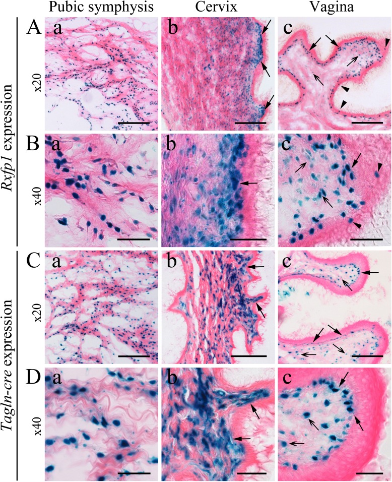
FIG. 2.
Rxfp1 expression and Tagln-cre-mediated recombination of ROSA26 reporter in reproductive organs of late pregnant females. A and B) Representative images of β-galactosidase staining of Rfxp1LacZ/+ female reproductive organs at Day 18.5 of pregnancy. Blue staining labeled by arrows or arrowheads shows gene expression. Aa and Ba) All cartilage cells of the pubic symphysis are strongly positive. Ab and Bb) In the cervix there is strong staining in the lamina propria (arrows) and stromal smooth muscle cells. Ac and Bc) In the vagina there is strong staining in the lamina propria (arrows) and in a few epithelial cells (arrowheads), and some weak staining in the vaginal stroma (arrows with open arrowheads) is shown. C and D) Representative images of β-galactosidase staining of ROSA26-LacZ, Tagln-cre female reproductive organs at Day 18.5 of pregnancy. Ca and Da) Intense staining is located in cells of the pubic symphysis. Smooth muscle cells of the cervix (Cb and Db) and vaginal stromal cells (arrows with open arrowheads; Cc and Dc) are strongly positive, as are the cells of the lamina propria (arrows). At least three mice of each genotype were analyzed. Bars = 100 μm (A and C) and 50 μm (B and D).

