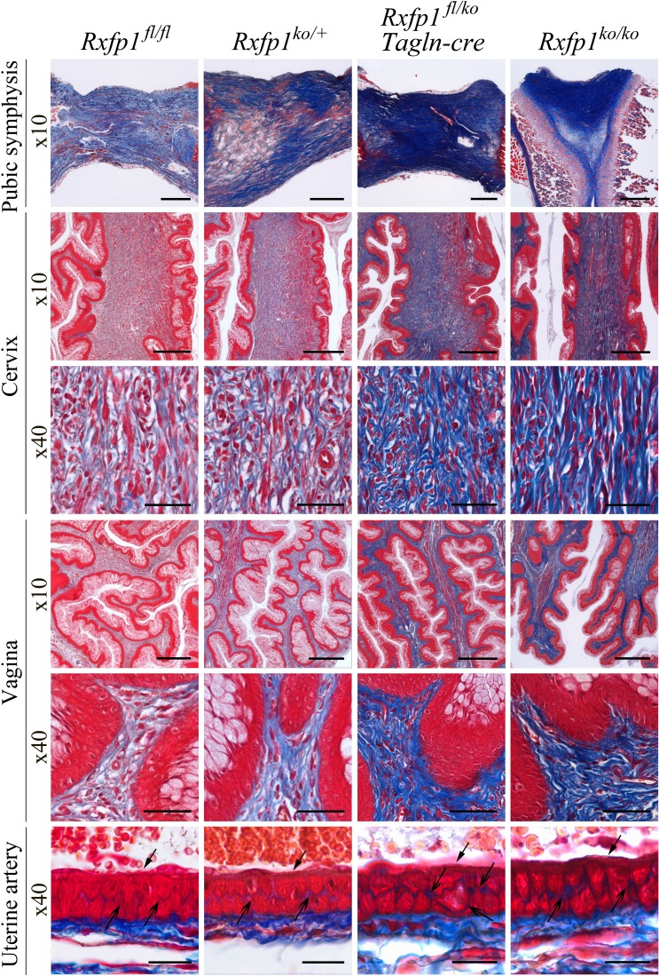
FIG. 6.
Increased collagen density in late pregnant females with global and smooth muscle-specific deletions of the Rxfp1 gene. Masson trichrome staining was used to visualize collagen organization within the cervix, vagina, pubic symphysis, and uterine artery wall. In all tissues from females with conditional and global Rxfp1 gene deletion, collagen staining is stronger and collagen fibers are less organized than in wild-type or heterozygous females. In the uterine artery the arrows show the more intense collagen staining within the vascular smooth muscle cell layer. Arrows with the open arrowhead show endothelial cells. Shown are representative images of at least three animals analyzed at ×10 and ×40 magnifications. Bar = 200 μm (for ×10) and 50 μm (for ×40).

