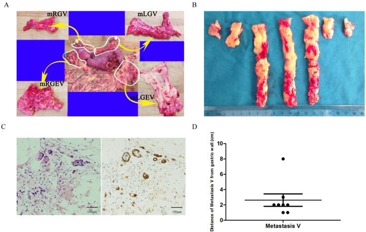
Fig 1. Detection of Metastasis V in gastric cancer patients.
(A) Large cross sectional tissue samples analysis of mesogastrium from surgically resected specimens. mLGEV, mRGEV, mLGV and mRGV were analyzed. (B) Continuous sections at 1-cm-width intervals of mesogastrium specimens. (C) Isolated cancer cells were detected in the mesogastrium of resected gastric cancer specimens by both HE staining (left) and immunohistochemistry with CK AE1/AE3 antibody (right). (D) Distance of Metastasis V from the gastric walls.

