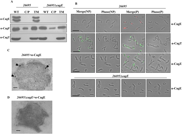
Fig 1. Sub-cellular localisation of CagE.
(A) Western blots showing sub-cellular localisation of CagE in wild-type Hp (26695) and 26695ΔcagE cells. WT, C/P, and TM indicate whole cell lysate, soluble (cytoplasmic/periplasmic) and total membrane fractions respectively. Western blots were probed with anti-CagE, anti-CagF and anti-CagT antibodies as indicated. (B) IFM showing CagE in wild-type Hp (26695) and 26695ΔcagE cells under permeabilised (P) and non-permeabilised (NP) conditions. Cells were probed with the anti-CagE, anti-CagT and anti-CagZ antibodies as indicated. CagT was used as a control for surface exposed proteins and CagZ was used as a control for inner membrane proteins. 26695ΔcagE cells were used as a negative control for anti-CagE antibody. Alexa fluor 594 (red colour) and Alexa fluor 488 (green colour) conjugated secondary antibodies were used to visualise the signals. [Out of 500 cells having fluorescent foci tested 100 foci were detected at the poles, 220 were at the middle and remaining 180 foci were detected near the poles]. Scale bars indicate 5 μm. (C) TEM showing inner membrane association of CagE in wild-type Hp (26695). (D) 26695ΔcagE cells stained with anti-CagE antibody. Cells were grown on BHI agar plates and immunogold labelling of ultrathin sections were performed as described in the materials and methods. Wild-type Hp 26695 and 26695ΔcagE (negative control) cell sections were probed with anti-CagE antibody and gold-labelled secondary antibody. Scale bars indicate 100 nm. Arrows indicate the location of gold-labelled CagE.

