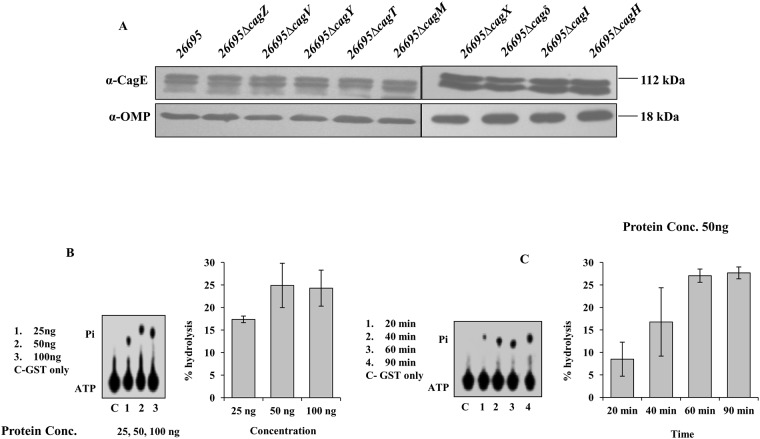
Fig 2. Western blots showing stability of CagE in several isogenic Cag mutants of Hp 26695 and ATPase activity of the CTD of CagE.
(A) Western blots showing stability of CagE in indicated strains. Antibodies used in Western blotting are indicated. OMP was used as loading control. (B) Concentration-dependent ATPase activity of the purified GST-CagEC (541–983, aa). Lane C: reaction with GST only; lanes 1–3: reactions with increasing concentrations of the protein as indicated. Positions of ATP and released Pi are indicated. (C) Time-dependent ATPase activity of the purified GST-CagEC (541–983, aa). Lane C: reaction with GST only; lanes 1–4: reactions with increasing time as indicated. Positions of ATP and Pi are indicated. Percent hydrolysis of ATP was plotted against protein concentration or against incubation time. Statistical error bars are indicated.

