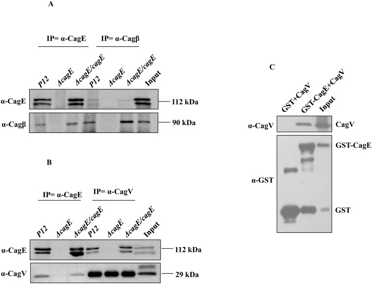
Fig 3. CagE interacts with Cagβ and CagV.
(A) Western blots showing co-immunoprecipitation (Co-IP) of Cagβ and CagE from cell extracts prepared from wild-type Hp (P12) and P12ΔcagE/cagE by anti-CagE and anti-Cagβ antibodies respectively. P12ΔcagE strain was used as a negative control. Antibodies used in the Western blots are marked. (B) Western blots showing Co-IP of CagV and CagE from cell extracts prepared from wild-type Hp (P12) and P12ΔcagE/cagE by anti-CagE and anti-CagV antibodies respectively. P12ΔcagE strain was used as a negative control. Antibodies used in the Western blots are marked. (C) Western blots showing GST-pull-down of CagV by GST-CagE. Antibodies used in Western blotting and precipitated proteins are indicated.

