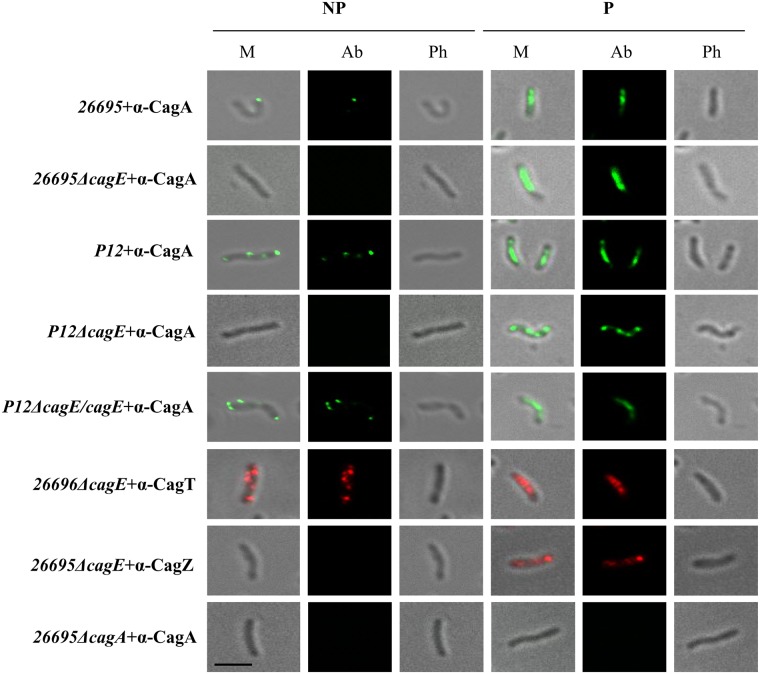
Fig 6. IFM showing CagE dependant surface localisation of CagA in wild-type Hp 26695 and P12 cells.
26695ΔcagA and P12ΔcagE strains were used as negative controls. Localisation of CagT and CagZ in 26695ΔcagE strains were used as controls for surface and inner membrane localised proteins respectively. NP and P stand for non-permeabilised and permeabilised cells respectively. M, Ab, and Ph stand for Merge, respective antibody used, and Phase respectively. Primary antibodies used are indicated. Alexa fluor 488 (green) and Alexa fluor 594 (red) conjugated secondary antibodies were used for immuno detection. Scale bar indicates 5 μm.

