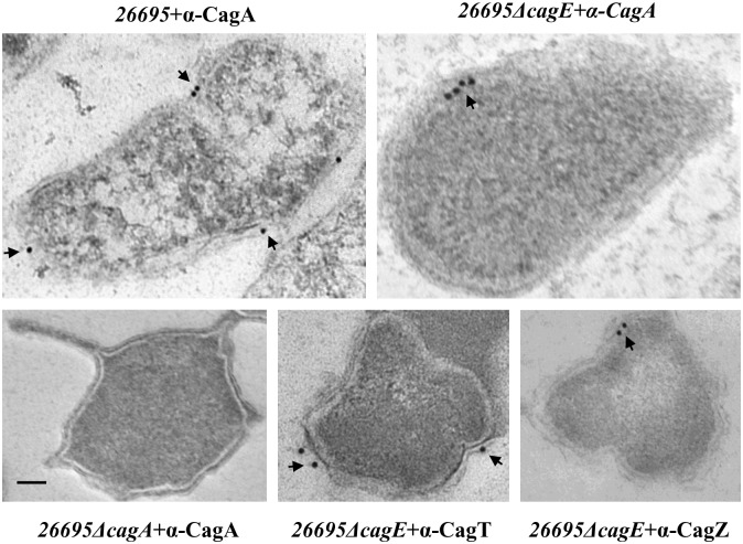
Fig 7. Immunogold electron microscopy showing localisation of CagA in wild-type and 26695ΔcagE strains.
Ultra thin sections of wild-type, 26695ΔcagE and 26695ΔcagA mutant cells were immunostained with anti-CagA and gold-labelled secondary antibody. 26695ΔcagA strain was used as a negative control for CagA antibody. Localisation of CagT and CagZ were used as a control for surface exposed and inner membrane proteins. Scale bar indicates 100 nm. Arrowheads indicate location of gold-labelled secondary antibody.

