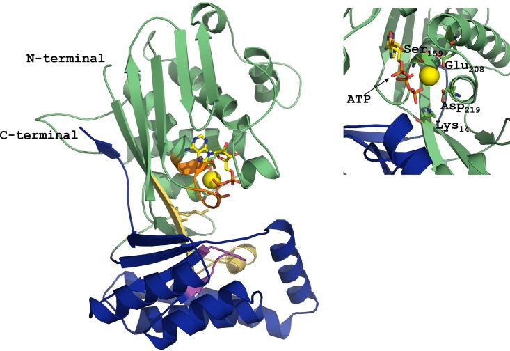
Fig 2. Overall structure of the A. aegypti MVK in complex with MgATP.
The N-terminal domain is shown in green and the C-terminal domain is shown in blue. Motif I (light yellow), Motif II (orange) and Motif III (magenta) are shown in different colors. Key amino acids (Lys14, Ser159, Glu208 and Asp219) are shown in the core of the protein. ATP and Mg (yellow ball) are also shown. The model was generated using the rat MVK (PDB code 1KVK) as template, using PyMOL.

