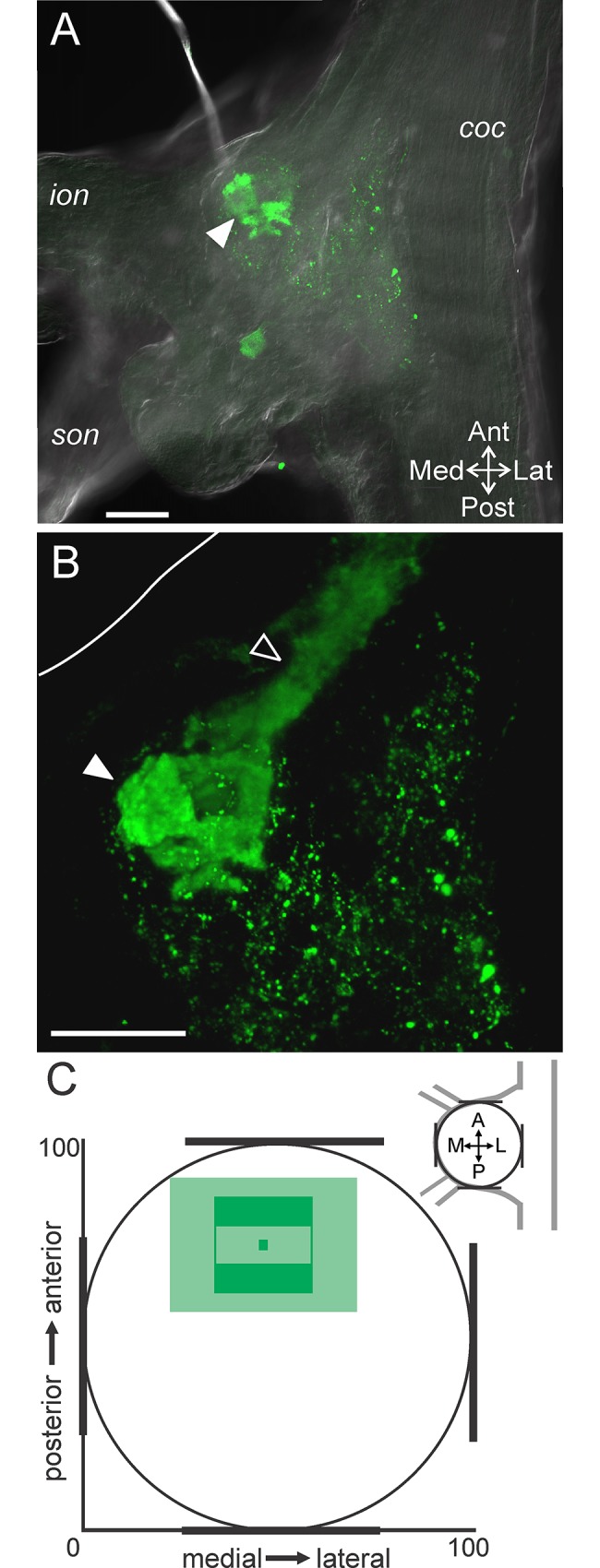
Fig 4. The ACO consistently occurs in the anterior region of the CoG.

(A) A single optical slice including fluorescence signal (green: CabTRP Ia-IR) and DIC optics demonstrates that the ACO (filled arrowhead) was located in the anterior region of the CoG. (B) A volume rendering (170 slices, 1.0 μm interval) of the same ganglion as in (A) illustrates that the POC axons (open arrowhead) entered the CoG from the anterior coc and terminated as the large ACO structure (filled arrowhead). White line marks the edge of the CoG visible in (A). Scale bars: 100 μm. (C) The average location of the ACO indicates a consistent occurrence in the anterior portion but a variable location across the mediolateral axis. The ACO location is reported as the average (dark green center box) and standard deviation (lighter green center box) of the center of the ACO and the average (dark green filled outer box) and standard deviation (lighter green outer box) of the spread of the arborization as measured vertically and horizontally from center. coc, circumoesophageal connective; ion, inferior oesophageal nerve; son, superior oesophageal nerve.
