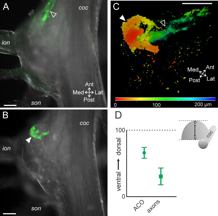
Fig 5. The POC axons and the ACO neuroendocrine organ differ in their dorsoventral locations.
Single optical slices with CabTRP Ia-IR (green) and DIC optics illustrate that (A) the POC axons (open arrowhead) entered the CoG near the ventral surface (70 μm from the ventral CoG surface), while (B) the ACO (filled arrowhead) occurred more dorsally (184 μm from ventral CoG surface, 94 μm from the dorsal CoG surface) than the POC axons. (C) Depth coding (red = dorsal, green/blue = ventral) applied to a volume rendering of a z-stack of the same CoG as above highlights that the POC axons (open arrowhead) are located more ventral than their termination as the ACO (filled arrowhead) (201 slices, 1.2 μm interval). (D) Average (± S.D.) locations of the center of the ACO structure (circle) and the POC axons (square) in the dorsoventral plane are plotted. Scale bars: 100 μm. coc, circumoesophageal connective; ion, inferior oesophageal nerve; son, superior oesophageal nerve.

