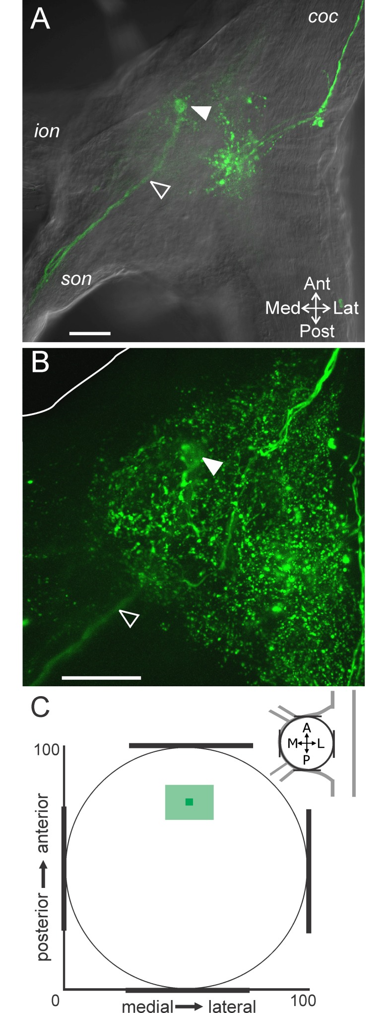
Fig 8. The GPR axon bundle projects into the anterior CoG region.
(A) The GPR axon bundle (serotonin-IR; open arrowhead) entered the ganglion through the son and projected into the anterior region of the CoG where the axons appeared to terminate in a compact bundle on this plane (filled arrowhead). The tissue was visualized with DIC optics (single optical slice). (B) A maximum intensity projection of the same CoG as in (A) reveals additional serotonin-IR while the GPR axon bundle is still evident projecting into the anterior region and appearing to terminate as a bundle (filled arrowhead) (268 slices, 0.8 μm steps). The white line indicates the outline of the tissue in the anteromedial region. Scale bars: 100 μm. (C) A plot of the average (± S.D.) location of the apparent GPR axon bundle termination indicates a consistent occurrence in the anterior CoG. coc, circumoesophageal connective; ion, inferior oesophageal nerve; son, superior oesophageal nerve.

