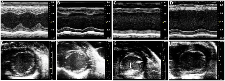
Fig 1. Representative M-mode (A-D) and short-axis views (E-H, at end diastole) following echocardiography of wildtype (A, E), early HF (B, F), acute HF (C, G), and chronic HF (D, H) mice.
M-mode images were obtained from a parasternal long-axis view to assess septal (top) and posterior (bottom) wall thickening. A mural echolucent mass on the posterior wall consistent with mural thrombus is seen (G, white arrow) as well as papillary muscle (G, grey arrow). Speckle within LV cavities represents spontaneous echo contrast. Scale bar is on the right axis (mm).

