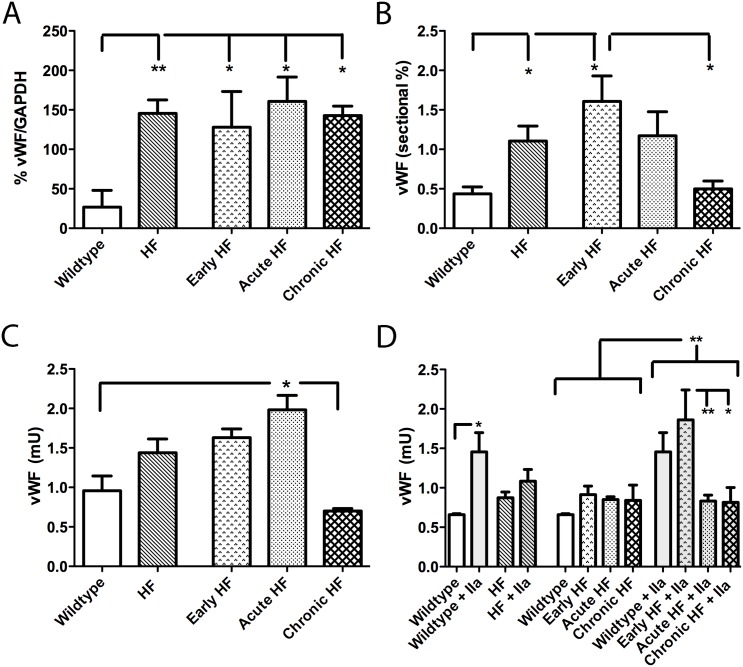
Fig 6. Quantitative determination of vWF: in plasma (A; immunoblot), by immunostaining in sectional endocardium (B; IHC), on the endocardial surface (C; ELISA), and following thrombin (IIa)-promoted vWF secretion from endocardium (D; ELISA).
Circulating vWF expression is higher in all groups of HF compared to wildtype mice (A). Sectional percentage of vWF staining is higher in early HF compared to both wildtype and chronic HF mice (B). Endocardial vWF protein levels are higher in acute HF compared to both wildtype and chronic HF mice (C). Thrombin-promoted vWF extrusion is higher overall than unstimulated endocardium (D). Thrombin-promotion of vWF extrusion is prominent in wildtype mice and in early HF mice (D). *p<0.05, **p<0.01.

