Figure 2.

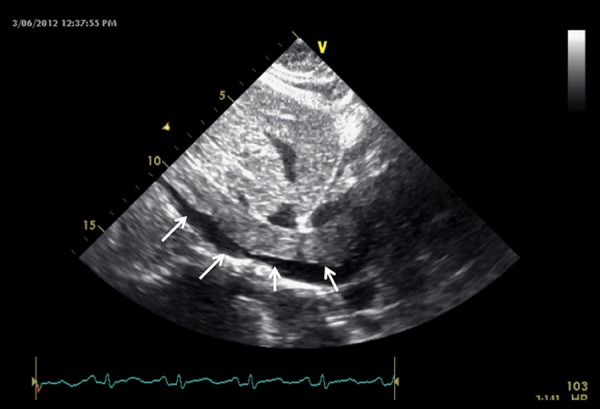
Transesophageal echocardiogram at the level of the IVC showing an elongated, tubular, worm-like echogenic mass within the lumen of the inferior vena cava (arrows).

Transesophageal echocardiogram at the level of the IVC showing an elongated, tubular, worm-like echogenic mass within the lumen of the inferior vena cava (arrows).