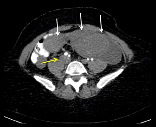
Figure 3.

Axial image of a contrast-enhanced abdomen and pelvis CT scan at the level of the lower abdomen. The enlarged uterus is visualized with multiple heterogeneous enhancing masses (likely leiomyomas) (white arrows). The right common iliac vein is enlarged, with intraluminal material of intermediate attenuation related to tumoral involvement (yellow arrow).
