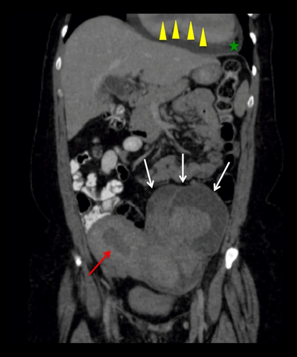
Figure 5.

Coronal reformat image of a contrast-enhanced Abdomen and Pelvis CT Scan. Enlarged myomatous uterus with multiple large masses (white arrows) showing heterogeneous enhancement in this contrast enhanced image, probably secondary to cystic degeneration of myomas. There is small amount of fluid within the endometrial cavity (red arrow). A moderately sized pericardial effusion is present (green star). Partially visualized tubular shaped filling defect within the right heart chambers, consistent with tumoral extension (yellow arrowheads).
